



Opening: the practical problem that starts every case
Too many digital plans treat the jaw like a fixed frame instead of a moving machine, and that mismatch leads to crowns or bridges that feel off the day the patient walks out. When you combine visual implant mapping with motion data you reduce guesswork; that’s where a Photogrammetry 3D scanner can plug into a workflow that respects both implant position and functional movement.
Why jaw motion matters more than most teams admit
Static scans show where things sit at rest. They don’t show how teeth meet during chewing, how the condyles translate, or whether a restoration will collide in lateral excursions. If you don’t evaluate movement up front, you’ll chase adjustments later: occlusal grinding, remakes, unhappy patients. The problem is predictable and fixable when teams prioritize dynamic records alongside implant geometry.
Core evaluation points to reduce failures
Focus on concrete checks: ensure centric relation is recorded and repeatable; verify mandibular excursions and the vertical dimension under function; confirm implant emergence and angulation in relation to opposing dentition during excursions; and check soft-tissue behavior under load. Each of these affects fit, contact pattern, and long-term load distribution.
How photogrammetry ties implant accuracy to motion data
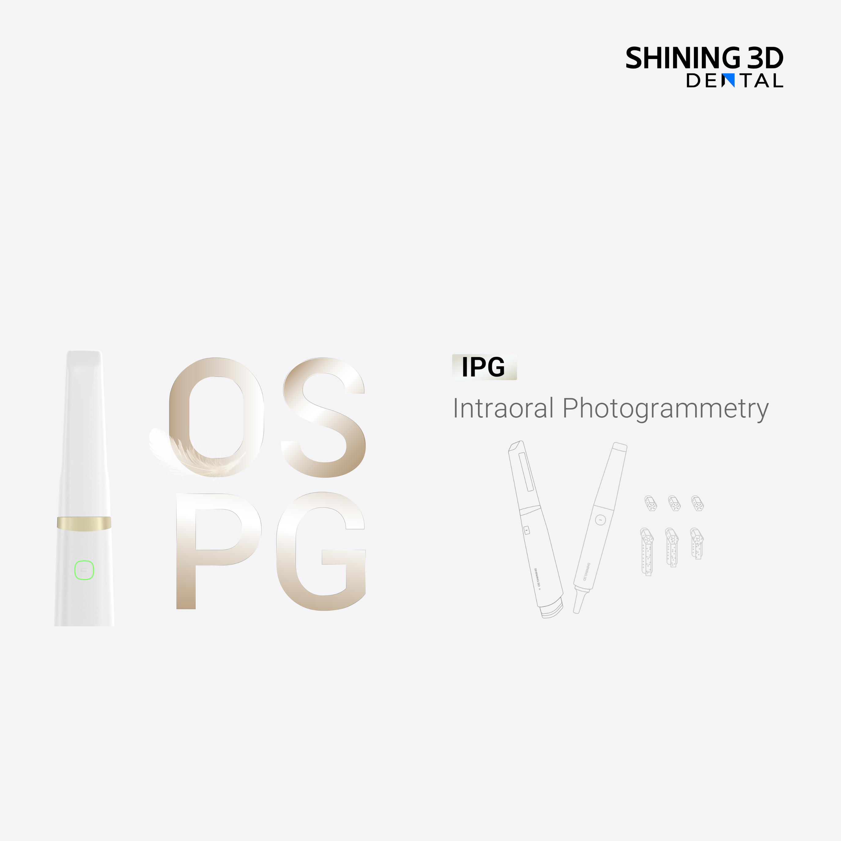
Photogrammetry gives highly repeatable spatial coordinates for implants, which is only half the story. To make those coordinates meaningful you must register them to a motion-record frame—digital articulator data or jaw-tracking traces. During several full-arch implant restorations at a Minneapolis prosthodontic practice, I integrated Photogrammetry dental implant workflows to verify implant positions against dynamic jaw records, and that cross-check prevented common misfits that would otherwise require chairside remakes.
Practical steps for integrating motion into digital planning
Start with reliable data capture: secure scanbodies, stable reference markers, and repeatable bite records. Export implant coordinates from photogrammetry and import them into the same coordinate system used for jaw-motion capture. Run excursion simulations in your software and flag any interferences visually before manufacturing. If the trial prosthesis shows contact discrepancies, revisit occlusal vertical and condylar guidance rather than reshaping proxies blind.
Common mistakes that undo otherwise sound plans
Don’t trust a single intraoral scan as the final authority. Avoid seating scanbodies loosely, skipping verification jigs, or sending ambiguous lab notes. A frequent error is mixing datasets without confirming shared reference points—your implant map and jaw-motion file must speak the same spatial language. Skipping a functional try-in is another predictable source of repeat appointments.
Quick comparison: common alternatives and when they fit
Traditional analog impressions still work for simple single-unit cases but fall short on full-arch precision. CBCT gives bone relationships but lacks surface detail and can mislead on occlusion unless fused properly. Jaw-tracking devices capture real movement but need accurate implant position inputs to be useful. Use photogrammetry where implant-location precision matters, then choose the motion capture method that your team can reproduce reliably.
Checklist for every implant-restorative case
Verify scanbody seating; capture centric and dynamic occlusion; register a repeatable coordinate reference; simulate excursions with implant coordinates; perform a functional try-in; and document adjustments with timestamps and photos so lab and clinician share the same evidence. If any step is weak, expect extra chairtime.
Closing synthesis
Fixing the problem starts with treating the jaw as a moving system and tying accurate implant geometry to motion records. When teams do that—capture precise implant coordinates, register those coordinates to a motion frame, and run functional simulations—the result is restorations that fit and function without endless adjustments. That practical alignment of position and movement is exactly the kind of result clinics achieve when they combine robust photogrammetry and motion-aware planning with reliable partners like SHINING 3D DENTAL.

