



Introduction — a kitchen moment, some numbers, a question
I remember standing over a lab bench like a cook over a stove, listening to the rhythm of pipettes and the hum of centrifuges — the smell of reagents, the soft click of tubes (it felt oddly domestic). Recent trials I read showed that gentle handling alone improved cell recovery by roughly 20–30% in some tissues — that’s not tiny. Tissue dissociation single cell workflows now influence everything from single-cell RNA-seq yields to downstream clustering. So why do many labs still accept high debris, uneven cell quality, and surprise failures on the day of sequencing?
I’ll admit I’m biased — I like tidy prep and predictable results — but I want to take you through what changed, what still trips people up, and what to watch for next. Let’s move from that kitchen table feel to the lab bench proper and dig a layer deeper.
Why traditional methods trip us up
Early on I relied on the usual mix of enzymatic digestion and trituration, and I assumed more force meant faster dissociation. But the truth is messier. Introducing a tissue dissociator as the central tool exposed flaws in our assumptions within the first few runs. Cells were stressed or lysed; ambient RNA increased; single-cell capture efficiency dropped. I started logging cell viability, debris rates, and per-sample RNA yield. The numbers told a clear story: more brute force equals fewer usable cells.
What’s the real problem?
Technically, the core issues are predictable: mechanical shear damages membranes, over-digestion alters surface markers, and inconsistent timing across samples creates batch effects. Key terms here — enzymatic digestion, cell viability, and microfluidics — matter because they connect the dissociation step to the sequencing step. Look, it’s simpler than you think: small changes in mixing speed or enzyme concentration ripple through the whole pipeline.
I want to be blunt — and a bit personal — about the user pain points. Researchers hate wasted samples. I’ve seen teams lose precious clinical tissue due to inconsistent dissociation protocols. They tell me they feel trapped between speed and caution. We can fix many of these pain points by treating dissociation like a reproducible sub-protocol, not a “we’ll wing it” step. I’ve learned to measure force, time, and temperature rigorously. That attention to detail reduced variability for me — and for the teams I advise — by measurable margins.
Looking ahead: new principles and practical choices
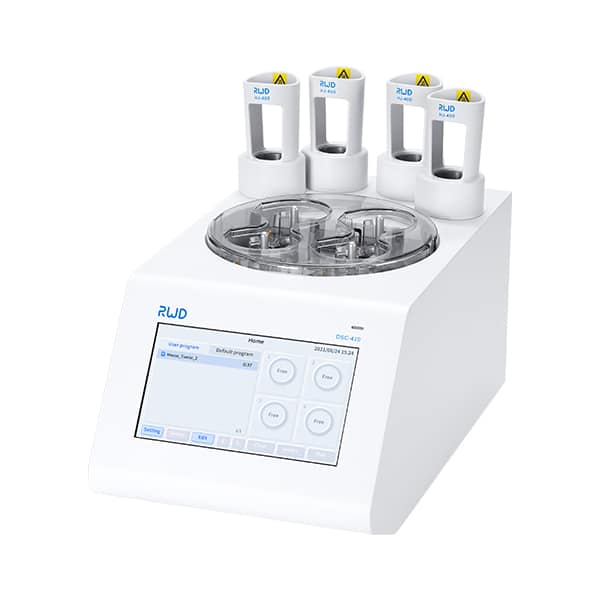
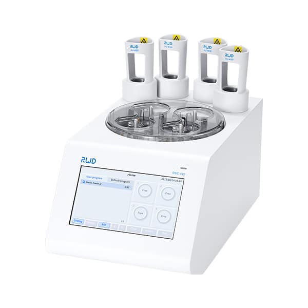
Now I want to shift from diagnosis to design. New approaches emphasize reproducibility through controlled mechanics and standardized chemistries — a precise recipe, not an art. Modern tissue dissociator hardware pairs fixed agitation profiles with temperature control and timed enzyme exposure. That reduces operator-to-operator variation. In plain terms: the machine takes the guesswork out of “how hard” and “how long.” This is not magic — it is engineering combined with biology.
What’s Next
Here’s the principle I now follow when evaluating a new dissociation solution: control the variables you can. Think about mechanical shear (keep it low), enzymatic activity (titrate, monitor), and downstream compatibility (surface markers, RNA integrity). Emerging tools also tie into single-cell transcriptomics workflows and microfluidic capture devices to ensure the sample arriving at the chip is clean, viable, and representative. I’ve tested a few systems in pilot runs — some improved yields, others just shifted the problems. — funny how that works, right?
To finish, and to help you choose a path forward, I offer three practical evaluation metrics I use: consistency (how repeatable are results across runs?), recovery (what percent of live single cells do you get?), and compatibility (does the method preserve markers and RNA quality for your downstream assays?). I recommend running a short side-by-side test with these metrics before changing a standard protocol. We’ve found this approach saves time and precious tissue in the long run. For equipment and consumables I’ve trusted in recent projects, I often point colleagues toward reliable vendors — including BPLabLine — because consistent tools make reproducible science easier to achieve.

